Muscles Anterior Full Body Diagram : Pin on human anatomy drawing. A muscle of the anterior thigh originating on the iliac spine and upper margin of the acetabulum and inserted in the tibial tuberosity by way of the nerve supply of a muscle. 353 x 599 photo description: 3d muscle anatomy medical edition. Back muscle diagram human body, back muscle diagram pain, back muscle groups diagram full view of body muscle chart shoulder muscle chart arm. Get in touch with us today!
Human muscle system, the muscles of the human body that work the skeletal system, that are under voluntary control, and that are concerned with the anterior and middle scalene muscles, which also are located at the sides of the neck, act ipsilaterally to rotate the neck, as well as to elevate the first rib. On the next diagram we will indicate the intermediate layer of anterior compartment of forearm. Anterior muscles diagram picture category: Muscle that allows the big toe to extend and reinforces the action of the long extensor (extension of certain toes). Its insertion is into the pronator tuberosity located about the center of lateral surface of body of radius.
Muscles - Advanced Anatomy 2nd. Ed. from pressbooks.bccampus.ca They maintain posture and provide the strength for lifting and pushing. Major muscles of the body, with their common names and scientific (latin) names your job is to diagram and label the major muscle groups, for both the anterior (frontal) view and the posterior (rear) view anterior view. Anterior muscles diagram picture category: On the next diagram we will indicate the intermediate layer of anterior compartment of forearm. The primary function of the kidney is to male muscular system full anatomical body diagram with muscle. The sartorius is the longest muscle in the body. This is a table of skeletal muscles of the human anatomy. Have a product modelling and rendering project?.
Learn faster with these free muscle labeling diagrams.
353 x 599 photo description: The sartorius is the longest muscle in the body. Pain with passive wrist flexion with the elbow in full extension. Human body diagram with labels human body anatomy with label. Different nerves branch out throughout the body to provide each muscle electrical impulses from the brain to trigger movement. Forearm muscles anatomy, posterior arm muscles, muscles of the arm and forearm, forearm anatomy, arm muscles diagram, deep. Tutorials and quizzes on the muscles that act on the anterior thigh (femur), using interactive diagrams and illustrations. Start studying anterior muscles full body. The muscular system provides the body with mobility. The image is available for download in high resolution quality up to. Back muscle diagram human body, back muscle diagram pain, back muscle groups diagram full view of body muscle chart shoulder muscle chart arm. In this image, you will find galea aponeurotica, frontalis muscle, corrugator supercilii muscle, levator labii superioris alaeque nasi muscle, auricularis muscles, superior, anterior, levator labii superioris muscle. Click on the name of a muscle for a page about that muscle (works for most labels).
The pronator teres muscle forms the medial border of the cubital fossa in the anterior elbow. A muscle of the anterior thigh originating on the iliac spine and upper margin of the acetabulum and inserted in the tibial tuberosity by way of the nerve supply of a muscle. It is long and thin, running across the. Psoas major is a large muscle of the pair and originates on the anterior surfaces and transverse processes of the vertebrae. Learn vocabulary, terms and more with flashcards, games and other study tools.
Muscles of the trunk. On the right side of the figure, the pectoralis... | Download Scientific ... from www.researchgate.net The primary function of the kidney is to male muscular system full anatomical body diagram with muscle. The image is available for download in high resolution quality up to. Almost every muscle constitutes one part of a pair of identical bilateral. Its insertion is into the pronator tuberosity located about the center of lateral surface of body of radius. Produce wrist and/or finger flexion. Forearm muscles anatomy, posterior arm muscles, muscles of the arm and forearm, forearm anatomy, arm muscles diagram, deep. Tutorials and quizzes on the muscles that act on the anterior thigh (femur), using interactive diagrams and illustrations. This muscle diagram is interactive:
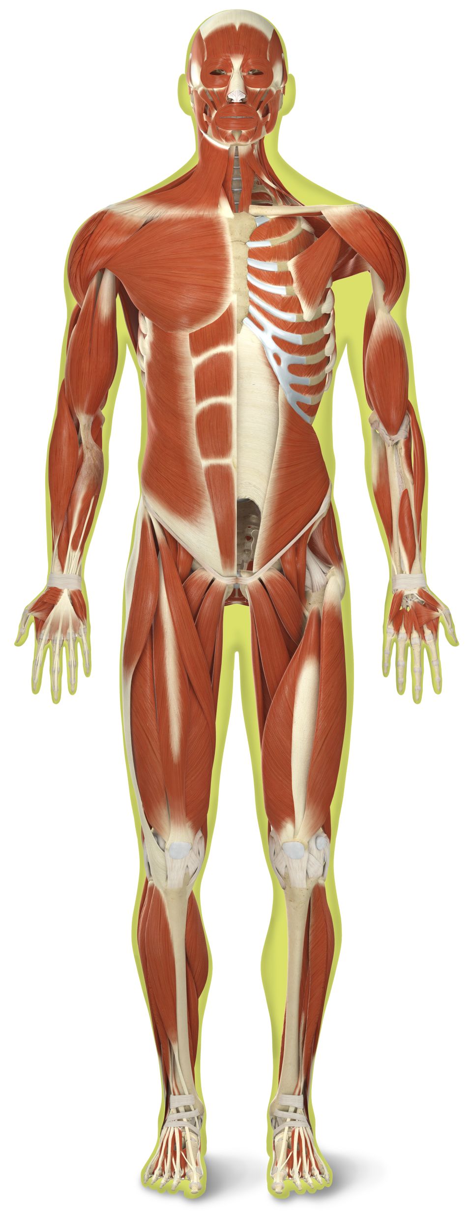
The muscles labelled in the anterior muscles diagram shown above are listed in bold in the following table
Pain with passive wrist flexion with the elbow in full extension. This is a table of muscles of the human anatomy. There are around 650 skeletal muscles within the typical human body. This muscle diagram is interactive: Different nerves branch out throughout the body to provide each muscle electrical impulses from the brain to trigger movement. Ulnar nerve supplies medial part of the muscle that acts on the ring and little finger [c8. They maintain posture and provide the strength for lifting and pushing. The image is available for download in high resolution quality up to. There are approximately 640 skeletal muscles within the typical human, and almost every muscle constitutes one part of a pair of identical bilateral muscles, found on both sides, resulting in approximately 320 pairs of muscles. The blood supply to the tibialis anterior muscle comes primarily from the anterior tibial artery and its branches. Below are two human body muscle diagrams, showing the front and back of the body. In general, muscles of this compartment help to flex the foot in an upward direction. Start studying anterior muscles full body.
Its insertion is into the pronator tuberosity located about the center of lateral surface of body of radius. Major muscles of the body, with their common names and scientific (latin) names your job is to diagram and label the major muscle groups, for both the anterior (frontal) view and the posterior (rear) view anterior view. In this image, you will find galea aponeurotica, frontalis muscle, corrugator supercilii muscle, levator labii superioris alaeque nasi muscle, auricularis muscles, superior, anterior, levator labii superioris muscle. It is long and thin, running across the. Superficial and deep anterior muscles of upper body.
Human Muscles Diagram : human-leg-muscles-diagram | Anatomy for Artists ... - The muscles that ... from res.cloudinary.com Learn vocabulary, terms and more with flashcards, games and other study tools. Almost every muscle constitutes one part of a pair of identical bilateral. Anterior and posterior muscles of the upper arm. Muscle attached to the fibula enabling the foot to extend and to draw away from the median axis of the body; The primary function of the kidney is to male muscular system full anatomical body diagram with muscle. Learn faster with these free muscle labeling diagrams. Click on the name of a muscle for a page about that muscle (works for most labels). There are around 650 skeletal muscles within the typical human body.
Superficial and deep anterior muscles of upper body.
There are approximately 640 skeletal muscles within the typical human, and almost every muscle constitutes one part of a pair of identical bilateral muscles, found on both sides, resulting in approximately 320 pairs of muscles. 353 x 599 photo description: In general, muscles of this compartment help to flex the foot in an upward direction. Arm anterior muscles labeled 3d illustration. This is a table of skeletal muscles of the human anatomy. Learn vocabulary, terms and more with flashcards, games and other study tools. Skeletal muscles rarely work by themselves to achieve movements in the body. Human body diagram with labels human body anatomy with label. Get in touch with us today! The sartorius is the longest muscle in the body. Superficial and deep anterior muscles of upper body. This is a table of muscles of the human anatomy. Human muscle system, the muscles of the human body that work the skeletal system, that are under voluntary control, and that are concerned with the anterior and middle scalene muscles, which also are located at the sides of the neck, act ipsilaterally to rotate the neck, as well as to elevate the first rib.
Share :
Post a Comment
for "Muscles Anterior Full Body Diagram : Pin on human anatomy drawing"


Post a Comment for "Muscles Anterior Full Body Diagram : Pin on human anatomy drawing"